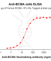
| Clone ID | DM6 |
|---|---|
| Target | |
| Synonyms | TNFRSF17 |
| Host Species | Rabbit |
| Description | Anti-BCMA antibody(DM6); Rabbit mAb |
| Delivery | In Stock |
| Uniprot ID | Q02223 |
| IgG type | Rabbit IgG |
| Clonality | Monoclonal |
| Reactivity | Human |
| Applications | ELISA; Flow Cyt; IF |
| Recommended Dilutions | Flow Cyt 1:100 |
| Purification | Purified from cell culture supernatant by affinity chromatography |
| Formulation & Reconstitution | Lyophilized from sterile PBS, pH 7.4. Normally 5 % – 8% trehalose is added as protectants before lyophilization. Please see Certificate of Analysis for specific instructions of reconstitution. |
| Storage & Shipping | Store at -20°C to -80°C for 12 months in lyophilized form. After reconstitution, if not intended for use within a month, aliquot and store at -80°C (Avoid repeated freezing and thawing). Lyophilized proteins are shipped at ambient temperature. |
| Background | The protein encoded by this gene is a member of the TNF-receptor superfamily. This receptor is preferentially expressed in mature B lymphocytes; and may be important for B cell development and autoimmune response. This receptor has been shown to specifically bind to the tumor necrosis factor (ligand) superfamily; member 13b (TNFSF13B:TALL-1:BAFF); and to lead to NF-kappaB and MAPK8:JNK activation. This receptor also binds to various TRAF family members; and thus may transduce signals for cell survival and proliferation. [provided by RefSeq; Jul 2008] |
| Usage | Research use only |
| Conjugate | Unconjugated |
| DIMA Disclaimer | All DIMA recombinant antibodies are genuinely generated by DIMA Biotech. They are all under patent application. Any protein sequencing or reverse engineering attempt is prohibited. We are actively scrutinizing all patent application to ensure no IP infringement. |
Anti-BCMA antibody(DM6); Rabbit mAb
Price: 10μg $99.00 ; 100 μg $438.00 ; 500 μg $1314.00
Product Data Dima FAQ
Images Dima FAQ
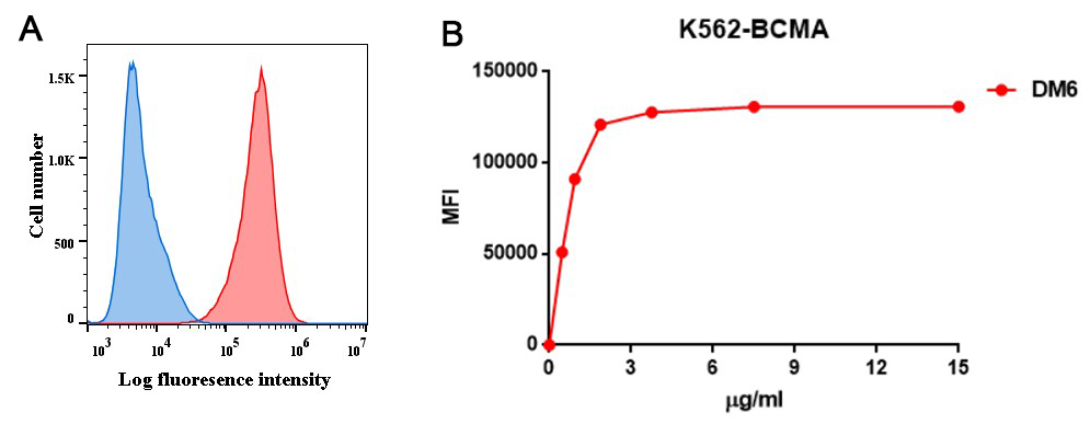
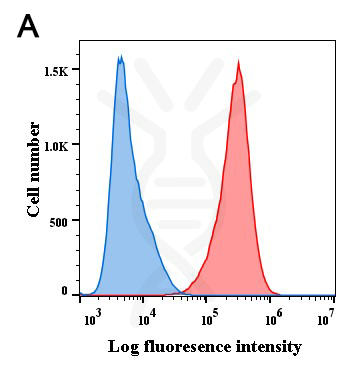
Figure 1. A. Flow cytometry analysis with anti-BCMA ( DM6) on K562-BCMA (Red histogram) (K562 cells stably transduced by human BCMA full length gene) and K562 (Negative control cell line) (Blue histogram). B. Flow cytometry data of serially titrated anti-BCMA ( DM6). The Y-axis represents the mean fluorescence intensity (MFI) while the X-axis represents the concentration of IgG used.
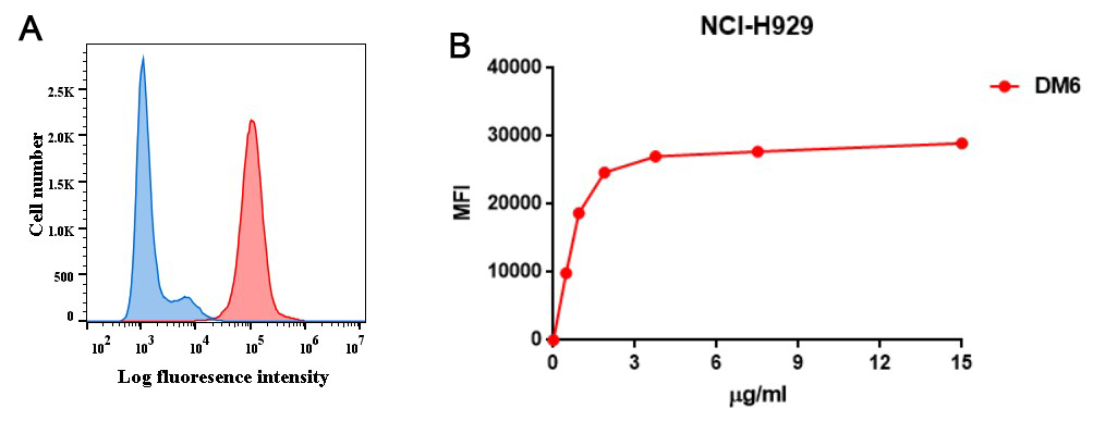
Figure 2. A. Flow cytometry analysis with anti-BCMA ( DM6) on NCI-H929 cells (Red histogram) or rabbit control antibody on NCI-H929 cells (Blue histogram). B. Flow cytometry data of serially titrated anti-BCMA ( DM6) on NCI-H929 cells. The Y-axis represents the mean fluorescence intensity (MFI) while the X-axis represents the concentration of IgG used.
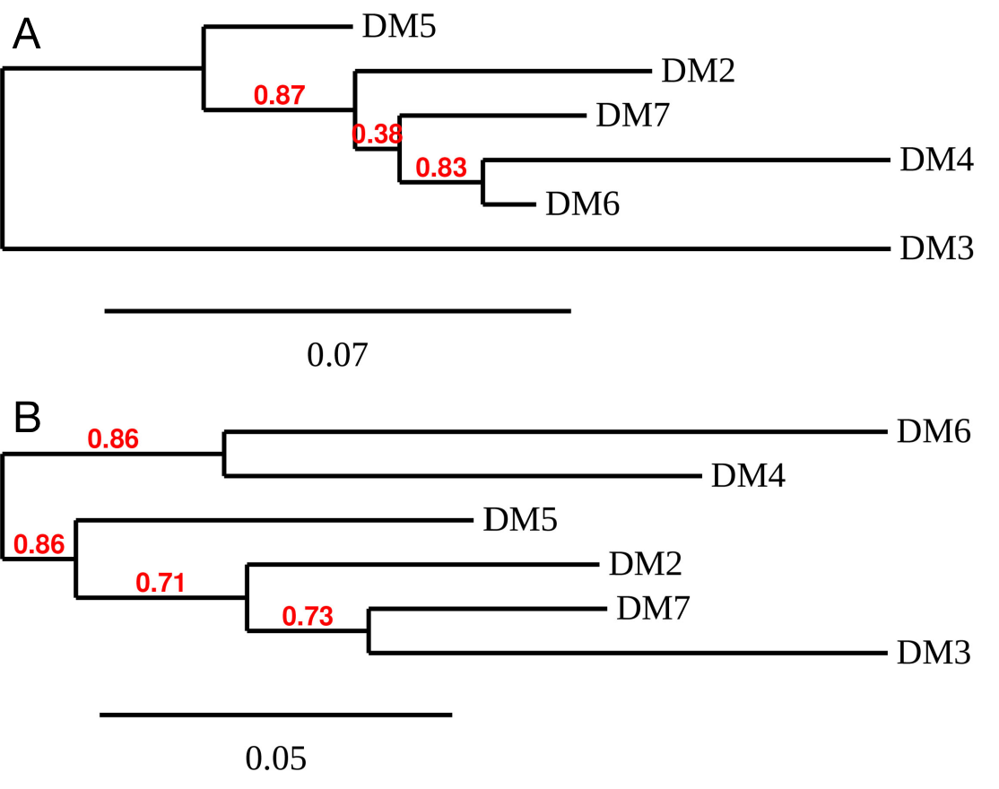
Figure 3. Phylogenetic analysis of different Anti-BCMA DimAb clones. A) heavy chain and B) Light chain.

Figure 4. Affinity ranking of different DimAb clones by titration of rabbit DimAb antibody concentration onto K562-BCMA or NCI-H929 cells. Different concentrations of various anti-BCMA DimAb clones were incubated with K562-BCMA ( A) or NCI-H929 cells ( B) at 4℃. Bound rabbit IgG was detected in flow cytometry analysis. The Y-axis represents the mean fluorescence intensity (MFI) while the X-axis represents the concentration of IgG used.
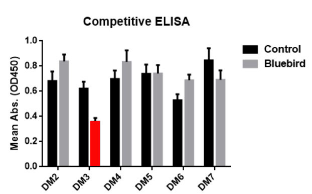
Figure 5. ELISA plate was coated with recombinant BCMA-hFc fusion protein PME100001 , followed by pre-blocking with huC11D5.3 antibody ( Grey bar) or rabbit control IgG ( Black bar), and then different rabbit DimAbs antibodies were added to check the competitive inhibition of huC11D5.3. DM3 clone exhibits the strongest inhibition ( Red bar). This data indicated that DM3 bind to the same epitope as bb2121.
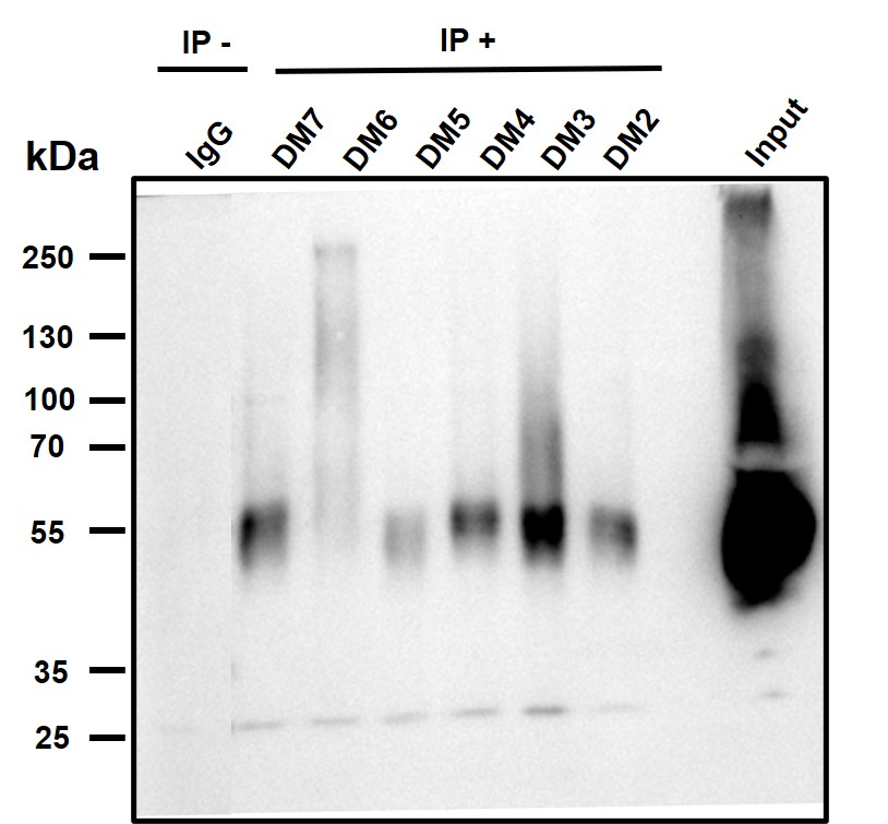
Figure 6. Immunoprecipitation analysis. Cellular overexpression lysates (made from HEK293F cells transfected with FLAG tagged human BCMA full length gene) were pre-incubated with 6 different rabbit DimAb clones and negative control IgG. The immunocomplexes were further pulled down by protein A beads, fractionated, and blotted with mouse anti-FLAG monoclonal antibody.
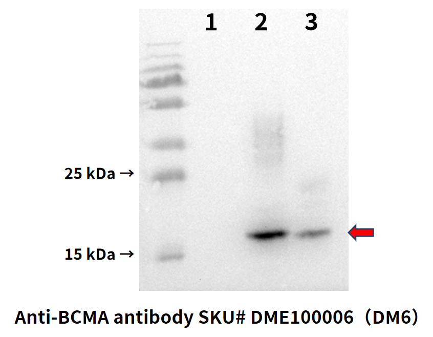
Figure 7. Western blot analysis of BCMA protein using Anti-BCMA antibody (Cat. DME100006) at 1:1000 dilution.
Secondary antibody: HRP Goat Anti-Rabbit IgG (H L) at 1:5000 dilution.
1. K562 cell lysate (negative control); 2. K562 cell lysate with overexpressed human BCMA proteins; 3. MM.1S cell lysate (native BCMA protein)
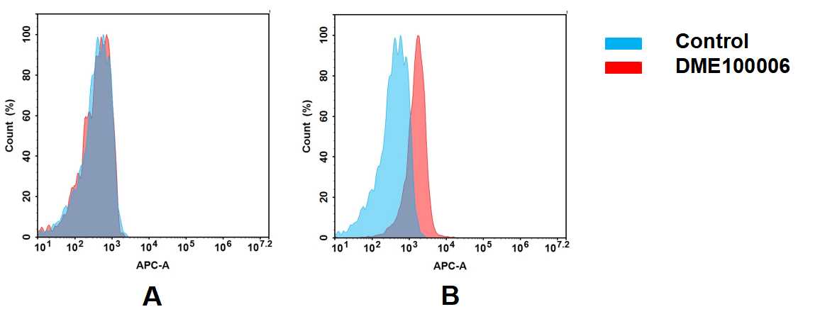
Figure 8. Flow cytometry analysis of antigen binding of rabbit anti-human BCMA mAb(DME100006).
(A) DME100006 does not bind to Jurkat cells that do not express BCMA.
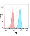
(B) A clear peak shift of DME100006 was seen compared to the control when incubated with BCMA-expressing MM.1S cells, indicating strong binding of DME100006 to BCMA. Antibodies were incubated at 2 μg/mL.
Related Products
ECD Proteins
SKU: PME101370 Target: BCMA
Price: 10μg $75.00; 50μg $259.00 ; 100μg $389.00
Biosimilar reference antibodies
SKU: BME100028 Target: BCMA
Application: ELISA; Flow Cyt
Price: 50μg $82.00 ; 100 μg $162.00
ECD Proteins
SKU: PME100001 Target: BCMA
Price: 10μg $82.00; 50μg $320.00 ; 100 μg $480.00
Biosimilar reference antibodies
SKU: BME100016 Target: BCMA
Application: ELISA; Flow Cyt
Price: 50μg $82.00 ; 100 μg $162.00
Biosimilar reference antibodies
SKU: BME100028B Target: BCMA
Application: ELISA; Flow Cyt
Price: 50μg $99.00 ; 100 μg $199.00

